
The short version
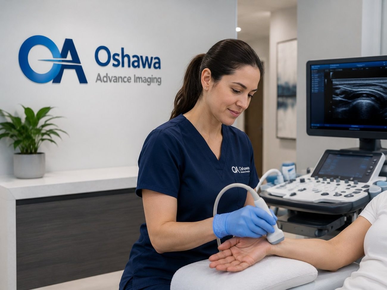
- Musculoskeletal ultrasound, often shortened to MSK ultrasound, images soft tissue: tendons, ligaments, muscles, nerves, bursae and joint linings.
- It is the only common imaging test that works dynamically, meaning the sonographer can scan while you move the joint.
- For full thickness rotator cuff tears, published studies put ultrasound accuracy in the same range as MRI. For subtle partial tears and surgical planning, MRI remains superior.
- No radiation, no enclosed tube, no problem with metal implants.
- You need a requisition from your physician or nurse practitioner. Ask them specifically about musculoskeletal ultrasound.
Shoulder, elbow, wrist, hip, knee and ankle pain account for an enormous share of visits to family doctors. The frustrating part for patients is what happens next.
An X-ray gets ordered, it comes back showing healthy looking bone, and the pain is still there. That is not a failed test. It is a test that answered the wrong question.
X-rays are excellent at bone. They are close to useless at the soft tissue that actually causes most joint pain: the tendon that is frayed, the bursa that is inflamed, the nerve that is compressed, the small tear in a muscle that never quite healed. Musculoskeletal ultrasound was built for exactly that soft tissue, and it is available right here in downtown Oshawa.
What musculoskeletal ultrasound actually sees
An ultrasound probe sends high frequency sound waves into the body and listens to the echoes that bounce back off different tissues. Because tendons, muscle fibres, fat and fluid all reflect sound differently, the machine can build a detailed picture of the layers under your skin. Modern high resolution probes resolve structures down to a fraction of a millimetre near the surface, which is precisely where most tendon and nerve problems live.
In practice, an MSK ultrasound can demonstrate:
- Tendon damage. Thickening, fraying, calcification, partial tears and complete ruptures in the rotator cuff, Achilles, patellar and other tendons.
- Inflammation. Fluid in a bursa or tendon sheath, and increased blood flow within inflamed tissue, which colour Doppler makes visible.
- Nerve compression. Swelling of the median nerve at the wrist, the classic finding in carpal tunnel syndrome.
- Fluid collections. Joint effusions, Baker cysts behind the knee, ganglion cysts at the wrist, and haematomas after injury.
- Lumps and hernias. Whether a palpable lump is a cyst, a lipoma, a lymph node or something that needs further work up, and whether a bulge in the groin or abdominal wall is a true hernia.
- Muscle injury. Tears and scarring in the calf, hamstring, quadriceps and elsewhere.
The advantage nobody talks about: movement
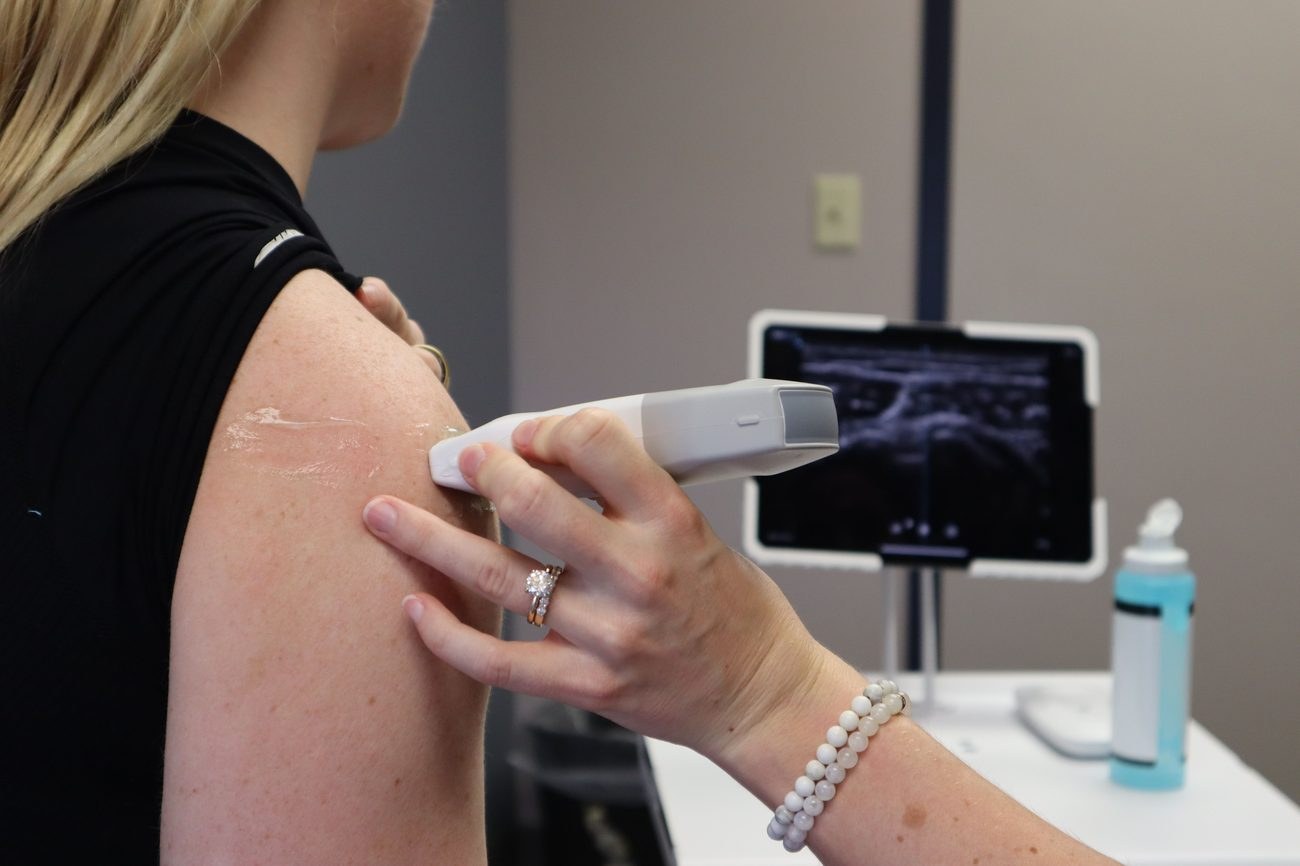
This is the part that surprises patients. An MRI takes a series of still pictures of a joint lying perfectly still inside a magnet. An ultrasound is live video. The sonographer can ask you to lift your arm, rotate your shoulder, flex your wrist or contract a muscle, and watch what happens in real time.
That matters because a lot of musculoskeletal pain is a movement problem, not a resting problem. A tendon that looks intact at rest may be seen catching against bone as the arm lifts. A hernia that disappears when you lie down often reappears the moment you stand and strain. A subluxing tendon at the ankle only misbehaves when the foot turns. Dynamic imaging catches these things; static imaging can miss them entirely.
The second underrated advantage is that the sonographer can ask you where it hurts and put the probe on that exact spot. Pointing at your pain and having someone image it directly is a surprisingly powerful diagnostic tool.
Operator matters. Ultrasound is more dependent on the skill of the person holding the probe than most imaging tests. That is why the study should be performed by trained professionals and interpreted by a qualified physician, and why the clinical detail on your requisition genuinely improves your result.
How ultrasound compares to MRI and X-ray
None of these tests is universally better. They answer different questions, and the right one depends on what your physician suspects.
Scroll sideways to see the full table.
| Ultrasound | MRI | X-ray | |
|---|---|---|---|
| Best at | Superficial soft tissue, tendons, nerves, fluid, lumps | Deep structures, cartilage, ligaments inside joints, bone marrow | Bone, fractures, alignment, arthritis |
| Radiation | None | None | Yes, low dose |
| Dynamic imaging | Yes, scans while you move | No | No |
| Metal implants | Not a problem | May be restricted, can cause artefact | Not a problem |
| Claustrophobia | Not an issue, open room | Can be difficult for some patients | Not an issue |
| Typical exam time | 20 to 45 minutes | 30 to 60 minutes | A few minutes |
| Compares both sides | Easily, in the same visit | Usually one side only | Possible |
What the evidence says about rotator cuff tears
Shoulder pain is the most common reason patients are sent to us for musculoskeletal imaging, so it is worth looking at the research honestly.
Multiple published studies comparing ultrasound and MRI against surgical findings have reported that full thickness rotator cuff tears are identified with broadly comparable accuracy by both methods, with sensitivity and specificity for ultrasound commonly reported in the high eighties to mid nineties when performed by experienced operators. Because ultrasound is faster, cheaper, better tolerated and easier to access, several of these studies conclude it is a reasonable first line investigation for suspected rotator cuff tears.
The honest caveat is equally well documented. Ultrasound is less reliable for subtle partial thickness tears, and MRI gives more consistent measurements of tear size, tendon retraction, muscle atrophy and fatty infiltration, all of which surgeons need when planning a repair. The literature also notes that where ultrasound is negative but clinical suspicion of a tear remains high, MRI is the appropriate next step.
How the two tests are usually sequenced
A simplified view of a common pathway for shoulder pain. Your physician decides the actual route.
Conditions we are commonly asked to image
Shoulder
Rotator cuff tendinopathy and tears, subacromial bursitis, calcific tendinitis, biceps tendon problems, and impingement assessed dynamically as the arm elevates.
Elbow
Tennis elbow and golfer's elbow, which are tendon problems at the outer and inner elbow, plus olecranon bursitis and ulnar nerve assessment.
Wrist and hand
Carpal tunnel syndrome, De Quervain tenosynovitis at the thumb side of the wrist, trigger finger, ganglion cysts, and tendon injuries. Wrist and hand imaging is one of the areas where the fine resolution of ultrasound is at its most useful.
Hip, knee and ankle
Greater trochanteric pain and gluteal tendinopathy at the hip, patellar tendinopathy and Baker cysts at the knee, and Achilles tendinopathy, plantar fasciitis and peroneal tendon problems at the ankle and foot.
Anywhere on the body
Soft tissue lumps, swellings, suspected hernias, and post injury assessment of muscle tears. If you can point to it, we can usually image it.
What your appointment is like
There is no preparation for a musculoskeletal ultrasound. You do not need to fast and you do not need a full bladder. The only practical advice is to wear clothing that lets you expose the area easily. For a shoulder study, a t-shirt or vest is far more convenient than a fitted dress.
During the exam you will sit or lie in a position that gives the sonographer access to the joint. Warm gel is applied and the probe is moved over the area, often repeatedly and from several angles, because tendons must be imaged in two planes to avoid a false appearance caused by the angle of the beam. You will likely be asked to move the joint, and you may be asked to point to the exact spot where the pain is worst. The unaffected side is sometimes scanned for comparison.
Most musculoskeletal studies take 20 to 45 minutes. Nothing is injected, there is no recovery period, and you can drive yourself home and return to work immediately.
How to get referred in Oshawa and Durham Region
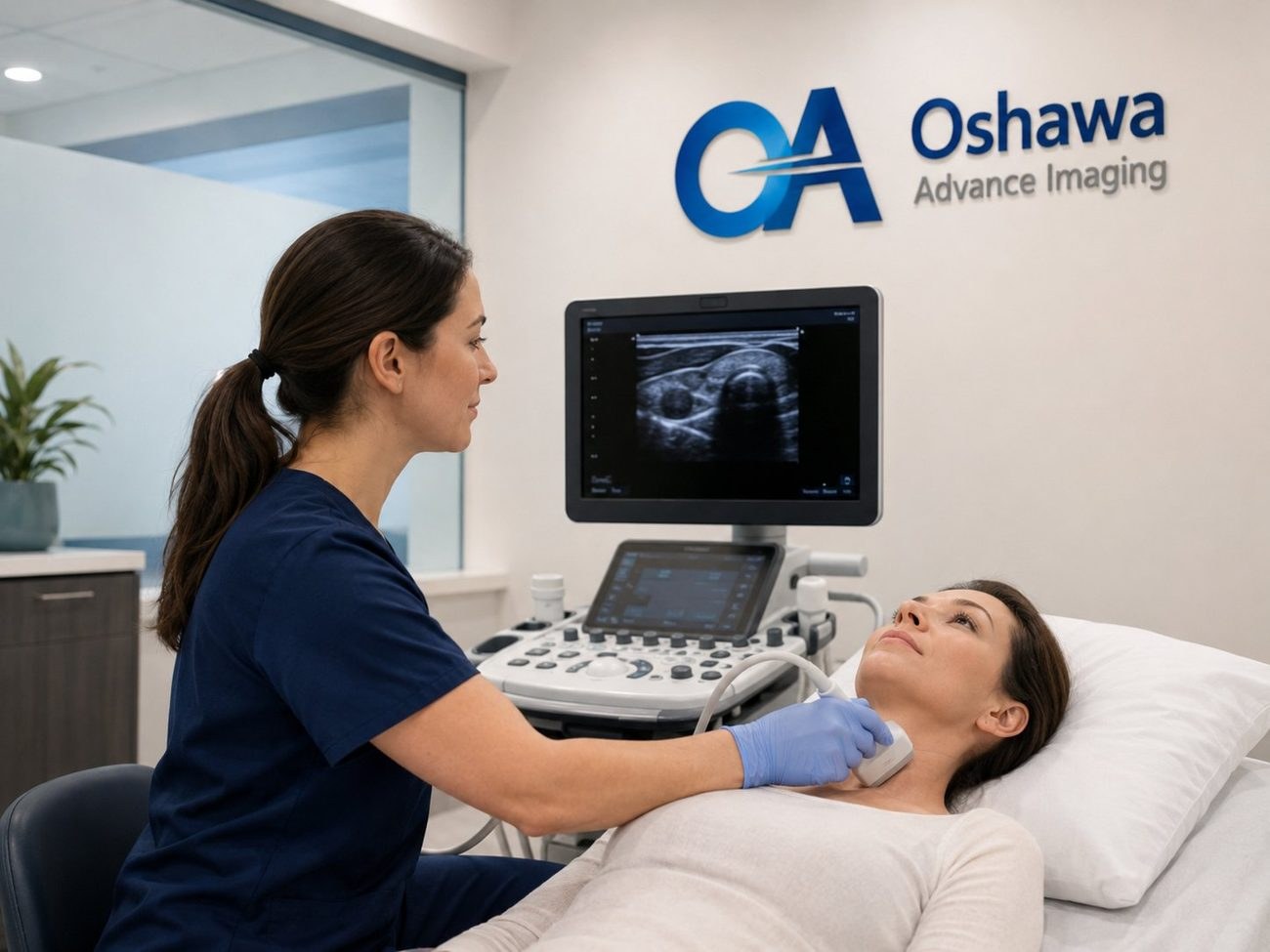
Diagnostic ultrasound in Ontario requires a requisition from a licensed physician or nurse practitioner, and an insured exam is covered when you have a valid Ontario health card and the study is medically necessary. Here is how to make that conversation productive:
- Be specific about the pain
Where exactly, how long, what makes it worse, and what movement you can no longer do. Duration and mechanism of injury genuinely change what the sonographer looks for.
- Ask directly about musculoskeletal ultrasound
Not every clinic in the region offers it, and some patients are told imaging is unavailable when what is meant is that it is unavailable locally. We offer it six days a week.
- Ask your physician to name the structure of interest
"Left shoulder, query supraspinatus tear" is far more useful on a requisition than "shoulder pain."
- Book promptly
Call us at +1 (289) 481-0263 or request an appointment online.
Physicians can fax requisitions to +1 (289) 731-2606 or download our form from the referring physicians page. Reports are sent back to the referring physician.
Common questions
Do I need to stop taking my medication before an MSK ultrasound?
No. There is no preparation for a musculoskeletal ultrasound and no need to change medication. Continue as your physician has prescribed.
Can ultrasound see arthritis?
Ultrasound can show the soft tissue consequences of arthritis, such as joint fluid, synovial thickening and erosions at the joint margin, and it can show crystal deposits associated with gout. It cannot assess deep joint surfaces or bone the way an X-ray or MRI can, so it is often used alongside them rather than instead of them.
Will the ultrasound hurt if my joint is already painful?
Pressure over an inflamed tendon or bursa can be briefly uncomfortable. Tell the sonographer, who can lighten the pressure or change approach. Many patients find the moment the probe reproduces their exact pain to be reassuring, because it confirms the source has been found.
I have a metal plate or joint replacement. Can I still be scanned?
Yes. Metal hardware does not interfere with ultrasound the way it can with MRI, which is one reason ultrasound is useful for patients who have had previous surgery.
Can I get an MSK ultrasound without a doctor's referral?
No. A requisition from a physician or nurse practitioner is required for diagnostic imaging in Ontario. If you do not have a family doctor, a walk-in clinic provider can assess you and issue one when imaging is appropriate.
This article is general information only and does not replace medical advice. Diagnostic accuracy figures referenced here are drawn from published peer reviewed literature comparing ultrasound and MRI and describe study populations, not individual results. Please speak with your physician or healthcare provider about your own health concerns.